
This is Part 1 of a four-part 1840 series, written for dental hygienists and anyone who wants to understand the connection between the mouth and the rest of the body.
I want to begin at the moment something has already gone wrong.
A portion of the heart muscle is being injured because its oxygen supply cannot meet its needs. If that ischemia is severe and sustained, muscle cells die. That is the injury we are working backward from.
For the next four letters, I want to trace one common route to that event: a clot forming on an atherosclerotic plaque in a coronary artery. Clinicians call an infarction from this process a type 1 myocardial infarction. Other heart attacks follow other routes. Keeping that distinction gives us a clear place to begin. Clinical definition
Start in the muscle.
The heart is full of blood, yet its muscle still needs blood delivered through the coronary circulation. When that delivery falls sharply, the cells have less oxygen available to produce the energy they use to contract and maintain their internal chemistry.
As the energy supply falters, contraction becomes impaired. Acidity and the movement of ions across cell membranes change. Electrical activity can become unstable. The pumping and electrical problems overlap; this is a living tissue responding to an injury, with several processes unfolding at once. Experimental physiology
A heart attack can trigger a dangerous rhythm and cardiac arrest, but the terms describe different events. A heart attack involves ischemic injury to muscle. Cardiac arrest means the heart has stopped providing effective circulation. NHLBI explanation
Look at the electrical signal. An ECG records electrical activity from different views of the heart. In the illustration below, find the sharp QRS complex, then follow the tracing into the ST segment.
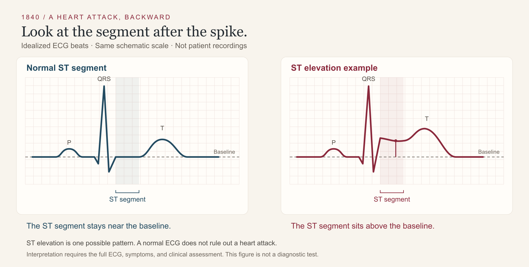
Figure 1. The shaded region identifies the ST segment. Elevation is one possible finding in an acute heart attack; some heart attacks have other changes or an initially normal ECG. Interpretation uses the full recording and clinical context. These are original teaching drawings, not patient recordings. Clinical guidance.
Now move backward into the artery.
In the pathway we are following, a clot has restricted the blood supply. It can grow rapidly on the surface of an existing plaque. The final loss of flow can therefore occur much faster than the underlying disease developed. NHLBI: causes
That clot contains platelets and fibrin. Platelets attach, activate, and recruit other platelets. Coagulation generates thrombin, which helps turn soluble fibrinogen into fibrin, a network that reinforces the clot.
To understand why that happened here, we have to go back one more step: to the surface on which the clot formed.
One possibility is plaque rupture. The fibrous cap over a plaque breaks, exposing material that promotes clotting.
Now look at actual tissue. In the right-hand column below, start with panel D and its boxed region. Panel E enlarges the junction between plaque tissue, marked with an asterisk, and thrombus, marked with a solid star.
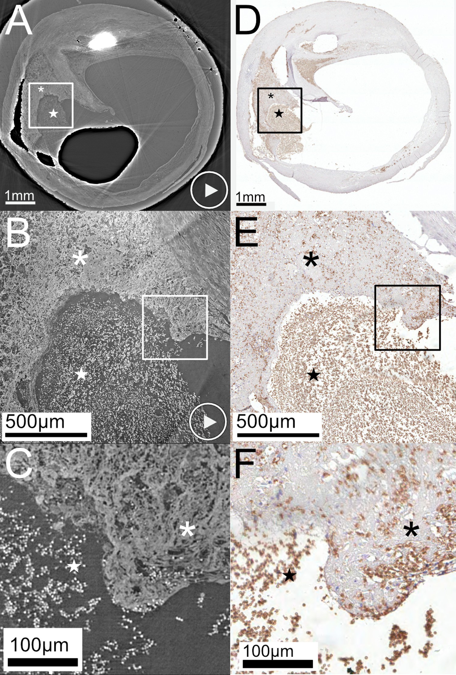
Figure 2. Human carotid-artery plaque with rupture and thrombus. The right column is microscopy; the left is synchrotron micro-CT. This specimen comes from the artery supplying the brain, and illustrates the structure of a rupture. It is not a coronary specimen or an image from the ECG patient. Original Figure 2, unchanged, from Truong et al., PLOS ONE, 2022, CC BY 4.0.
Another is plaque erosion. Injury at the surface allows a thrombus to develop without the same rupture through the cap. A calcified nodule is a third recognized substrate. Human coronary imaging has documented these different appearances. We should learn them before treating every coronary event as the same mechanical failure. Original OCT study
The forward sequence for the pathway we have just traced is:
Plaque-surface disruption → clot formation → reduced coronary flow → ischemic injury → infarction if the injury becomes irreversible.
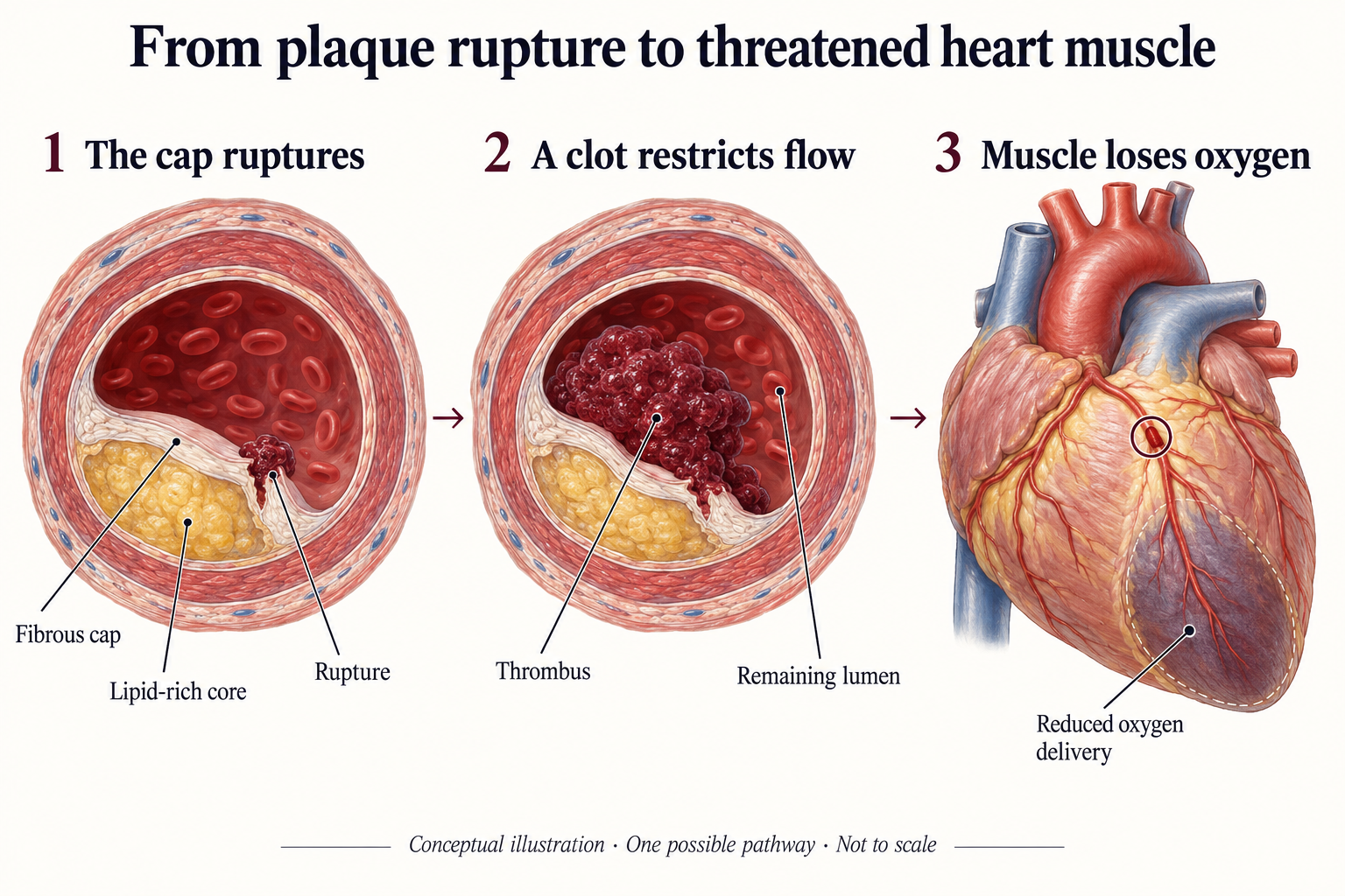
Figure 3. Follow the relationship between the plaque in the wall, the clot in the lumen, and the muscle downstream. This AI-assisted teaching illustration shows one possible pathway and is not to scale. The affected territory and severity vary between patients.
The timing and extent of injury vary. Rapidly restoring blood flow can preserve threatened muscle. That is why recognizing a possible heart attack and activating emergency care takes priority over finishing an examination or deciding whether discomfort is dental. NHLBI: treatment
For a hygienist, that distinction is practical.
Jaw discomfort can be part of a cardiac presentation. Chest pressure, shortness of breath, sweating, nausea, or discomfort extending into the arm, back, neck, or jaw deserve attention in context. A person does not need every symptom on the list. If a heart attack is suspected, call 911 and activate the office emergency response. NHLBI: symptoms
Between emergencies, there is a different window for our work. We can keep an accurate medical history, document changes, and communicate findings that need medical attention. We can also explain what we know without giving a patient a certainty the evidence does not support.
That includes the mouth–heart connection. Periodontal disease is associated with cardiovascular disease. Whether treating it prevents heart attacks remains unresolved. We will examine both the mechanisms and that evidentiary gap in the fourth letter. Updated AHA statement summary
For now, we have reached the plaque beneath the clot.
The next letter goes into the artery wall: what accumulated there, which cells arrived, what failed to clear, and how a lesion acquired the capacity to cause trouble.
— Thad
For the next team huddle: Trace the sequence from the injured muscle back to the plaque surface. Then locate the office emergency plan and confirm who calls 911, who retrieves the AED, and who directs the response.
Follow the four-part series
- Start with the clot. You are here: the injured muscle, the lost blood flow, and the plaque surface.
- What was in the artery before the clot? The plaque, the cells within it, and mineralization. Coming next.
- Pressure, flow, and mouth chemistry. Hypertension, nitric oxide, and the oral nitrate pathway. Forthcoming.
- Where does periodontal disease enter? Oral pathogens, inflammation, and what the evidence can and cannot establish. Forthcoming.
Subscribe to receive the remaining three parts. If you already receive 1840 by email, you are on the list.
If this helped, send it to one hygienist you work with. And tell me: which step would you like me to explain more closely? Email readers can reply to this letter; web readers can write to me. Please leave out identifying patient details.
Look closer: actual ECG recordings
These independent examples have different layouts and come from different people. They are not before-and-after recordings, and they are not from the microscopy specimen above.
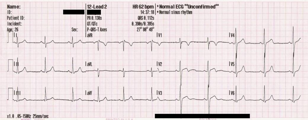
Reference recording: a 12-lead training ECG shared by MoodyGroove, public domain. The machine's normal interpretation is marked unconfirmed; this is a teaching reference, not independent clinical validation.

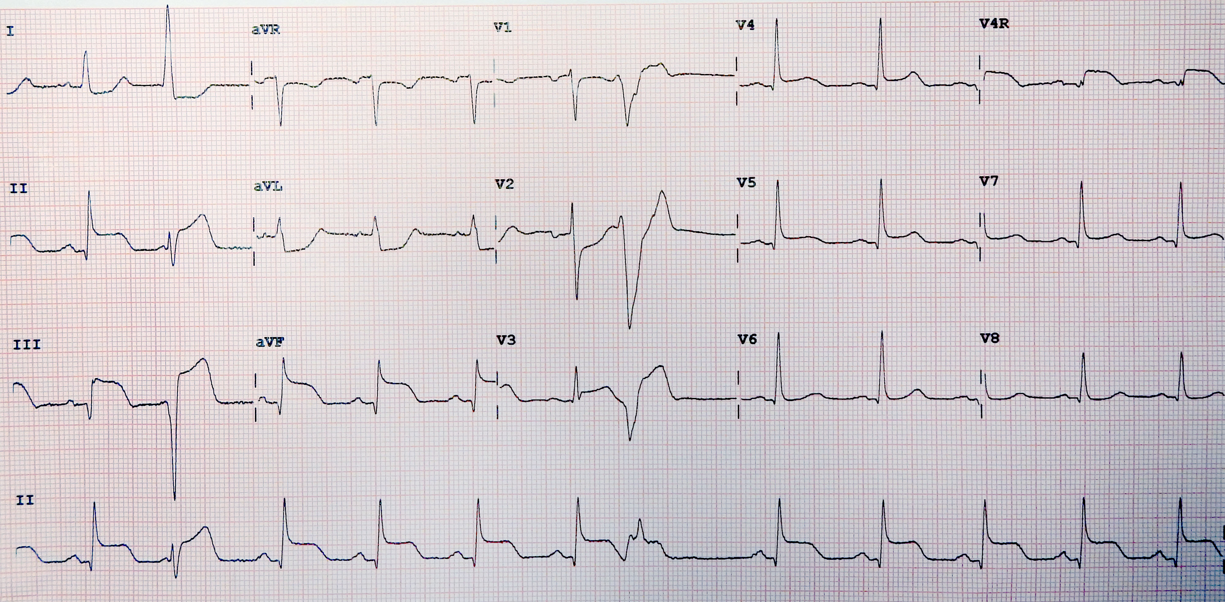
Infarction example: look at leads II, III, and aVF. The source identifies inferior and right-ventricular infarction. This 15-lead recording also includes additional views and a premature ventricular beat, so compare the ST segments rather than expecting identical overall shapes. Image: James Heilman, MD; derivative by Andrewmeyerson, source, CC BY-SA 4.0. Reproduced without further alteration.

For a coronary histology example, see Peter Anderson's ruptured coronary plaque with thrombus, UAB PEIR. This is linked for further study; it is not reproduced here.